
")

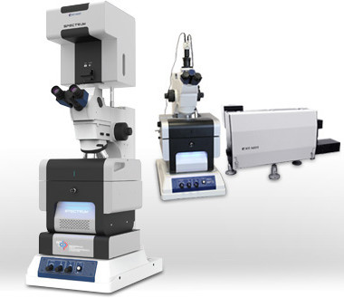
Sistema automatizzato AFM-Raman-SNOM per una ampia serie di applicazioni
Informazioni generali
Il primo strumento AFM-Raman-SNOM interamente automatizzato al mondo. È l'integrazione di un AFM con varie tecniche ottiche: microscopia confocale raman/a fluorescenza, SNOM, TERS, etc. Rende semplice una caratterizzazione comprensiva di un campione a livello nanometrico.
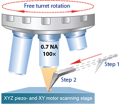
- Immagini AFM e Raman confocale simultanee usando diversi obiettivi (fino a 100x).
- Rotazione libera della torretta del microscopio (fino a quattro obiettivi) con la punta AFM sul campione.
- Rimozione automatica della punta AFM (per obiettivi con una breve distanza di lavoro o quando la punta non serve).
- HotSpot – localizzazione automatica della regione attiva TERS* sulla punta.
- Funzionamento interamente automatizzato e grande semplicità di utilizzo.
* Tip Enhanced Raman Scattering.
|
Due semplici passi per ottenere immagini Raman/Fluerescenza
Passo 2 Imamgini simultanee AFM e Raman/Fluorescenza dell'area selezionata |
 |
Features
The new SPECTRUM instrument was developed based on NT-MDT more than 15 years experience in building AFM - Raman – SNOM systems. The unique features include: full automation, advanced AFM capabilities, complete integration with optical techniques.
Unique integration of SPM with optics for AFM - Raman - SNOM – TERS
- Upright or Full Transmission configuration.
- Upright microscope with 4 position revolving turret. Possibility to install up to 4 different objectives starting from the lenses for large area visualization to high numerical aperture objectives for high optical resolution,
- Scanning by laser spot. This option is provided by very stable scanner-mirror with closed loop capacitance sensors. Latter allows to position the laser spot with a high precision on the tip apex.
- Fiber or direct input/output of the excitation laser/registered signal. Specially designed optical mechanical unit allows to input the incident laser by using high transmission optical fiber and output the collected signal through the fiber directly to the monochromator. Otherwise it is possible to couple directly SPECTRUM system with commercially available spectrometers (Solar, Renishaw).
- SNOM. Due to Full Transmission configuration it is possible to perform aperture and apertureless SNOM using cantilever or fiber probes
Automation
- Sample movement.
- Laser/cantilever/photodiode system alignment.
- Automatic removal of AFM probe (when low working distance objectives are used or when AFM probe is not required).
- Probe approach & retraction.
- Scanning parameters adjustment.
Unique SPM capabilities
- Low noise. Sample scanning with resolution down to atomic.
- Large sample size (up to 50mm×50mm). Special sample holder for slides (75mm×25mm).
- AFM, STM and tuning fork operation; measurements in liquid.
- More than 30 advanced SPM modes supported - together with Raman.
MultiScan
Sample survey with high resolution. Automated high resolution AFM - Raman imaging without limitations of the piezo-scanner range.
Automation
High-precision positioning motors allow to automate basic operations, including the adjustment of the OBD system, positioning of measuring heads and sample, etc.
Automated sample positioning (35×35 mm)*
High precision positioning motors equipped with optical sensors allow automated AFM-Raman imaging of any sample areas (within 35mm×35mm travel range).
Cantilever deflection system auto alignment
In several seconds get the laser aligned to the tip and photodiode position optimized. Special algorithms give a high precision and high speed of cantilever deflection system alignment.
Applications
- Graphene, carbon nanotubes and other carbon materials
- Semiconductor devices
- Nanotubes, nanowires, quantum dots and other nanoscale materials
- Polymers
- Optical device characterization: semiconductor lasers, optical fibers, waveguides, plasmonic devices
- Investigation of cellular tissue, DNA, viruses and other biological objects
- Chemical reaction control
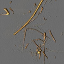
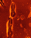
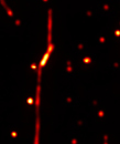
| Mo oxide nanowires, 5 ×5 µm |
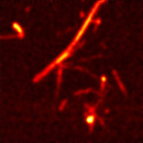
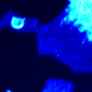
CdS nanowires, 4 ×4 µm |
|||
 |
 |
 |
 |
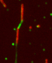 |
| AFM | Mo oxide Raman peak | AFM | Fluorescense image | Overlapped Raman image from CdS and PANI |
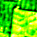
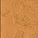
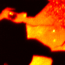
| Artificial diamond characterization, 2 ×2 µm | Graphene, 5 ×5 µm | |||
 |
 |
 |
 |
 |
| AFM topography | Raman mapping of intensity of 1333 1/cm band | AFM | Raman map of 2D-band | Raman map of G-band |
Specifications
Measuring modes and techniques
AFM (contact and amplitude modulation), AFM spectroscopy, AFM lithography (force, current, voltage), Raster Spring Imaging, Lateral Force Microscopy, Force Modulation Microscopy, Scanning Spreading Resistance Microscopy, Piezoresponse Force Microscopy and Switching Spectroscopy, EFM, Kelvin Probe Force Microscopy, MFM, STM (microscopy, spectroscopy, lithography).
Confocal Fluorescence microscopy, Confocal Raman microscopy, Confocal Rayleigh microscopy, TERS, TEFS, Wide field optical microscopy, SNOM (all types of probes, all modes)
Measuring heads
- AFM,STM and Tuning Fork
- Laser based cantilever deflection detection system automated adjustment and targeting
Sample
- Dimensions: up to 50/10 mm in diameter/height
- Sample weight: up to 2 kg
- Heating: from RT to 150 °С
Scanning system
- Scanning type: by sample
- Range: 100x100×10 μm (CL)
Resolution
- Noise XY: no more 0.3 nm (with closed loop sensors)
- Noise Z (RMS, 10-1000 Hz bandwidth): 0.06 nm (typical)
Sample positioning system
- Movement: automated, binded with the videomicroscope
- Range XY: 35x35 mm; 5×5 mm while using with the bottom objective
- Min. step: 0.35 μm
- Repositioning precision: 3 μm
Approach
- Automated approach by tip
- Range: 10 mm
Vibroisolation
- Active vibration isolation system
- Optical table (optional)
Optical parts
- Spectral range*: 450-1050 nm
- Spectral resolution: Depends on the grating and spectrometer used
- Objectives:
100x 0.7NA
10x 0.28NA
Any other objective (optional) - Detectors**:
TE cooled (down to -100 °C) CCD camera.
EMCCD camera is optional.
Photon multiplier (PMT) or avalanche
photodiode (APD) in photon counting mode or other type of CCD camera (UV, IR)
Photon multiplier for fast confocal laser
(Rayleigh) imaging - Confocal maps resolution***:
with the probe, for blue laser with 100x 0.7NA
XY: <400 nm
Z: <800 nm
without the probe, for blue laser with 100x 0.95NA
XY: <250 nm
Z: <500 nm - Bottom objective (optional)
Movement: motor and piezo (optional)
Range: 25 mm by motor, 100 μm by piezo
Min. step: 0.1 μm by motor, 3 nm by piezo
* Other spectral range available upon request.
**Depends on the system configuration